The following information and graphics was compiled from various WWW sources including the OFA website, discussion lists, and other sites.

During an OFA evaluation, there are 9 different anatomic areas of the hip that are evaluated (Figure 1).
1. Craniolateral acetabular rim : This should be sharp and clearly defined with no irregularities or spurring.
2. Cranial acetabular margin : Should be smooth and symmetrically curved
3. Femoral head (hip ball) : Should be spherical and convexity should smoothly fit the acetabular socket. A line drawn between point 1 and point 6 should encompass between 50% to 66% of the area defined by a line drawn between point 8 and point 6.
4. Fovea capitus (normal flattened area on hip ball) : Should be well-defined. A ligament called the ligamentum capitis connects point 4 to point 5 and serves to anchor the head in the socket and still permit rotational movement of the head within the socket.
5. Acetabular notch
6. Caudal acetabular rim : This should be sharp and clearly defined with no irregularities or spurring.
7. Dorsal acetabular margin : This should be sharp and clearly defined with no irregularities or spurring.
8. Junction of femoral head and neck : head and neck should be fused and the neck should not show any demineralization or evidence of weakness in the bone.
9. Trochanteric fossa : A line drawn down the midpoint of the femoral head (#3) to the midpoint of the trochanteric fossa (#9) should make a 130 degree angle with a line drawn down the midline of the long axis of the femoral shaft.
The radiologist is concerned with deviations in these structures from the breed normal. Congruency and confluence of the hip joint (degree of fit) are also considered which dictate the conformation differences within normal when there is an absence of radiographic findings consistent with HD. The radiologist will grade the hips with one of seven different physical (phenotypic) hip conformations: normal which includes excellent, good, or fair classifications, borderline or dysplastic which includes mild, moderate, or severe classifications.
Seven classifications are needed in order to establish heritability information (indexes) for a given breed of dog. Definition of these phenotypic classifications are as follows:
1. Excellent
2. Good
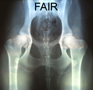
3. Fair
4. Borderline
5. Mild
6. Moderate
7. Severe
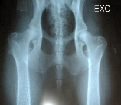
Now to analyze an x-ray that was rated OFA Excellent.
NOTE: Remember the dog is laying on it’s back when the x-rays are taken, so left is right and right is left.
 |
First check the positioning of the hips. To do this thoroughly, you need the full x-ray and not just the part that shows the hip joint. In the above photograph the alignment of the spine should be parallel to the two outer white lines. The ideal mid axis is shown in yellow while the actual axis is shown by the white. It is extremely difficult to get an ideal alignment. The above picture is about as close as most people will get. But when analyzing the film you have to first look at the alignment and adjust your interpretation based on the alignment. Another point is the horizontal yellow line across the tops of the greater trochanter of the femur must make 90 degree angles with the outer white lines. In this case, due to the slight misalignment, the horizontal yellow line slices just beneath the top of the left greater trochanter for it to make it’s 90 degree angle. This tells us the left hip will be slightly off, not because of the joint but because of the alignment. |
 |
Line A goes from the dorsal (upper) lip of the acetabulum to the ventral (lower) lip. This line when drawn shows that 66% or more of the femoral head is within the cup of the acetabulum (green line). This is especially so on the right side. On the left side the depth appears to be slightly less, but BEWARE! This is due to the very slight misalignment of the long axis of the animal. This is why doing the alignment test first is so important since it will throw the cup estimates off target. In this case the dogs left hip coverage is actually at least as much as the right. The angle between lines B & C are also measured since they represent a measure of the angulation of the femoral neck versus the shaft. In this case the hip has near perfect angulation. |
 |
Lastly the contour of the acetabular cup is looked at (Red Arrows) and the corresponding contour and fit of the femoral head is examined (Yellow arrows) No irregularities are seen and the shape and fit between cup and head is excellent. |
 |
Compare this to an x-ray rated by OFA: Moderate Dysplasia The differences are relatively clear between the two on every aspect after compensating for alignment. However, the alignment with the spine is off by quite a bit, it would not have hurt the owner to request another x-ray. |
The following x-rays were evaluated by OFA as indicated (click for larger view):